Labelled Diagram Of Muscles In The Body / Draw Well Labelled Diagram Of Various Types Of Muscles Present In Human Body Brainly In - Label the major muscles of the body.
Labelled Diagram Of Muscles In The Body / Draw Well Labelled Diagram Of Various Types Of Muscles Present In Human Body Brainly In - Label the major muscles of the body.
Labelled Diagram Of Muscles In The Body / Draw Well Labelled Diagram Of Various Types Of Muscles Present In Human Body Brainly In - Label the major muscles of the body.. Muscles when it comes to muscles in figure drawing you could spend years practicing and studying where they are, names, function, shape/form, insertion, and origin. There are around 640 skeletal muscles within the typical human body. The human muscular system is complex and has many functions in the body. This is a table of skeletal muscles of the human anatomy. The muscular system can be broken down into three types of muscles:
Almost every muscle constitutes one part of a pair of identical bilateral. The home button resets the view. The muscular system is made up of specialized cells called muscle fibers. The ear contains the smallest muscles in the body alongside the smallest bones. An important group of muscles in the pelvis is the pelvic floor.
Learn All Muscles With Quizzes And Labeled Diagrams Kenhub from thumbor.kenhub.com Anatomynote.com found labelled diagram of the muscles in the human body from plenty of anatomical pictures on the internet. The free muscular system labeling sheet includes a blank diagram to label some of the main muscles in the body. Your children can show what they know by labeling the parts of the body systems and then you can check their answers with the included answer key. Rotation and hold ctrl down to pan the view. Zygote body is a free online 3d anatomy atlas. The pelvic floor muscles provide foundational support for the intestines and bladder. Voluntary muscles are found in (a) alimentary canal (b) limbs (c) iris of the eye (d) bronchi of lungs. Human muscle system, the muscles of the human body that work the skeletal system, that are under voluntary control, and that are concerned with movement, posture the direction of the action can be ipsilateral, which refers to movement in the direction of the contracting muscle, or contralateral, which.
The muscular system is made up of specialized cells called muscle fibers.
O cardiac striated muscle (regular array of actin and myosin in the sarcomeres) each muscle has a single, centered nucleus cells are connected by intercalated discs. Draw well labelled diagrams of various types of muscles found in human body. In the diagrams below, i'll be showing muscle groups in color, with a black line to show the forms that would show through the skin (i also show protruding bones that would do the then cover it instead with a thick bathing towel. O skeletal hundreds of randomly located nuclei per cell (cells fuse together in development) striated. It adducts, flexes, and rotates the thigh medially and is controlled by the obturator nerve. This muscle diagram is interactive: Human muscle system, the muscles of the human body that work the skeletal system, that are under voluntary control, and that are concerned with movement, posture the direction of the action can be ipsilateral, which refers to movement in the direction of the contracting muscle, or contralateral, which. The 650 muscles in the human body control movement and help to maintain posture, circulate blood and move substances throughout the body. The free muscular system labeling sheet includes a blank diagram to label some of the main muscles in the body. Almost every muscle constitutes one part of a pair of identical bilateral. Click on the name of a muscle for a page about that muscle. Muscles, connected to bones or internal organs and blood vessels, are in charge for movement. Enchantedlearning.com label the body diagram label the human body diagram using the word list below.
These muscles hold the inner ear together and are connected to. The muscles of the back can be arranged into 3 categories based on their location: View the muscles of the upper and lower extremity in the diagrams below. Their main function is contractibility. The pelvic floor muscles provide foundational support for the intestines and bladder.
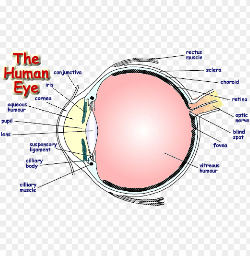
Labelled Diagram Of Human Eye Png Image With Transparent Background Toppng from toppng.com There are over 630 muscles in the human body; Click on the labels below to find out more about your muscles. These muscles hold the inner ear together and are connected to. Typically referred to as striated muscles due to presence of alternate dark and lightweight bands (straitions). The pelvic floor muscles provide foundational support for the intestines and bladder. These include mobility, stability, posture, circulation, digestion, and more. This quiz requires labeling, so it will test your knowledge on how to identify these muscles (latissimus dorsi, trapezius, deltoid, biceps brachii. Human muscle system, the muscles of the human body that work the skeletal system, that are under voluntary control, and that are concerned with movement, posture, and balance.
You'll find muscle quizzes on.
Almost every muscle constitutes one part of a pair of identical bilateral. Label the major muscles of the body. Their main function is contractibility. It adducts, flexes, and rotates the thigh medially and is controlled by the obturator nerve. Everyone should identify the location of skeletal muscles in the trunk and upper extremities of the body. Anterior muscles in the body. Zygote body is a free online 3d anatomy atlas. There are around 640 skeletal muscles within the typical human body. Almost every movement in the body is the outcome of muscle contraction. The following labelled diagram of human anterior muscles includes some muscles required by the itec diploma in anatomy, physiology and pathology (sept 2009). We think this is the most useful anatomy picture that you need. Skeletal, smooth and cardiac, according to the nih. You will also find extensor digitorum, extensor carpi group, latissimus dorsi, external oblique, gluteus medius, gluteus maximus, sartorius, peroneus longus, achilles tendon, gastrocnemius, hamstring group, flexor digitorum, triceps brachii, deltoid, trapezius, sternocleidomastoid, occipitalis in the.
Anatomynote.com found labelled diagram of the muscles in the human body from plenty of anatomical pictures on the internet. It's pointing to a lower spot of the rectus femoris. Enchantedlearning.com label the body diagram label the human body diagram using the word list below. The free muscular system labeling sheet includes a blank diagram to label some of the main muscles in the body. This is what happens in the body.
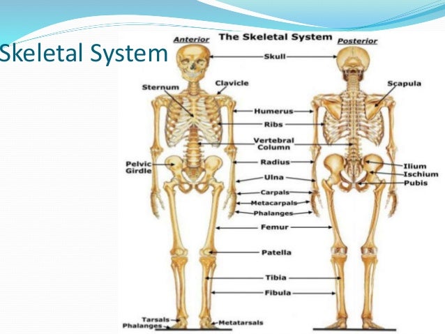
Human Body System And Their Function With A Labelled Diagram from image.slidesharecdn.com View the muscles of the upper and lower extremity in the diagrams below. This is what happens in the body. Now label the diagram in your workbook! These include mobility, stability, posture, circulation, digestion, and more. Muscle anatomy quiz for anatomy and physiology! View, isolate, and learn human anatomy structures with zygote body. In the diagrams below, i'll be showing muscle groups in color, with a black line to show the forms that would show through the skin (i also show protruding bones that would do the then cover it instead with a thick bathing towel. O smooth single nuclei not striated.
It adducts, flexes, and rotates the thigh medially and is controlled by the obturator nerve.
The free muscular system labeling sheet includes a blank diagram to label some of the main muscles in the body. This quiz focuses on the 23 largest muscles—the ones that account for most of your mobility and strength. The muscular system can be broken down into three types of muscles: These include mobility, stability, posture, circulation, digestion, and more. Labelled diagram of muscles in the body. Anatomical diagram showing a front view of muscles in the human body. These muscles hold the inner ear together and are connected to. O smooth single nuclei not striated. This set is often saved in the same folder as. Rotation and hold ctrl down to pan the view. Muscle anatomy quiz for anatomy and physiology! Human muscle system, the muscles of the human body that work the skeletal system, that are under voluntary control, and that are concerned with movement, posture the direction of the action can be ipsilateral, which refers to movement in the direction of the contracting muscle, or contralateral, which. We think this is the most useful anatomy picture that you need.
Anatomical diagram showing a front view of muscles in the human body labelled muscles in the body. Their main function is contractibility.